
Blood markers point to inflammation in 2 CMT types, study finds
CMT1A, CMT2A patients have higher levels of markers in blood
Written by |
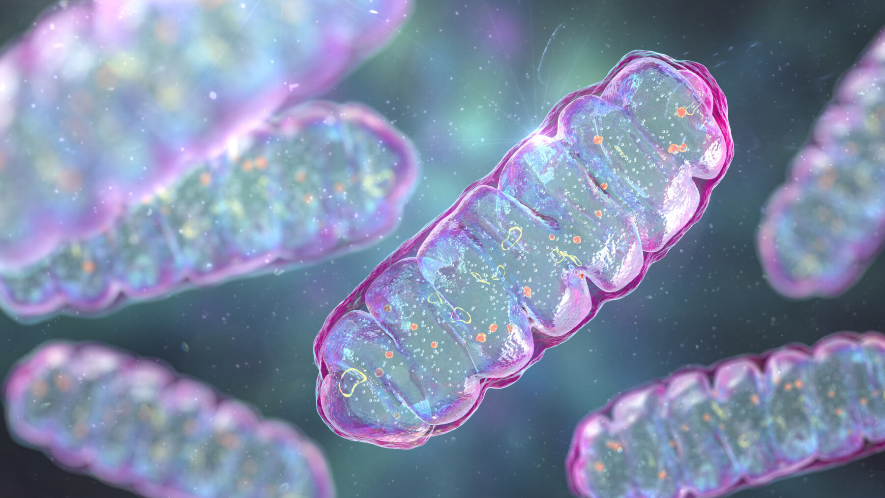
Mitochondria produce energy inside cells.
People with Charcot-Marie-Tooth disease types 1A (CMT1A) and 2A (CMT2A) have elevated blood levels of mitochondrial DNA and interleukin (IL)-1 beta, a molecule involved in inflammation, a study found.
“These findings suggest association between increased circulating mitochondrial DNA and inflammation in CMT disease,” the researchers wrote. “However, no subtype-specific differences were identified between CMT1A and CMT2A.”
Mitochondria are the structures that produce energy inside cells. While most of a cell’s genetic material is in its nucleus, mitochondria have their own DNA. In response to cellular damage, fragments of mitochondrial DNA (cell-free mtDNA) can reach the bloodstream and activate immune responses.
The study, “Circulating cell-free mitochondrial DNA and inflammation in Charcot-Marie-Tooth 1A and Charcot-Marie-Tooth 2A,” was published in Molecular Immunology.
CMT is a group of inherited disorders that affect the peripheral nerves, which connect the brain and spinal cord with the rest of the body. This results in progressive muscle weakness and CMT symptoms such as difficulty walking and reduced sensitivity to temperature or touch.

Relationship between mtDNA, inflammation markers
CMT1A and CMT2A affect peripheral nerves in different ways. CMT1A primarily involves damage to the protective coating of nerve fibers (myelin), while CMT2A affects the nerve fibers themselves. Mitochondrial dysfunction has also been implicated in CMT and related disorders.
To investigate the relationship between mtDNA in blood and markers of inflammation in CMT, researchers in China analyzed data from 51 people with CMT — 42 with CMT1A and nine with CMT2A — and 13 healthy controls. The mean age was 37.1 for participants with CMT1A and 24.9 among those with CMT2A. Overall, 60.8% of the CMT patients were men.
Those with CMT1A typically had muscle weakness and wasting in the limbs, particularly the legs, along with sensory loss. A higher proportion of those with CMT2A developed weakness earlier in life, with eight patients unable to walk before age 20.
Looking specifically at one mtDNA gene, ND1, and a nuclear DNA gene, ACTB, the levels of circulating cell-free mtDNA and the ratio of mitochondrial DNA to nuclear DNA were significantly higher in participants with CMT than in healthy controls. Nuclear DNA is the genetic material found in the cell nucleus. However, neither measure differed significantly between the two CMT groups.
Blood levels of IL-1 beta were also significantly higher in both CMT groups than in healthy controls and were significantly higher in participants with CMT2A than in those with CMT1A. However, there was no significant association between IL-1 beta and cell-free mtDNA in both CMT groups.
Biopsy samples of the sural nerve, a sensory nerve located in the back of the lower leg, were analyzed in 24 participants with CMT1A and five with CMT2A. Results revealed nerve fiber damage, structures called onion bulbs that can form when nerves undergo repeated cycles of myelin loss and repair, and mitochondrial degradation.
In participants with CMT2A, a higher mtDNA to nuclear DNA ratio was significantly associated with a higher number of onion bulbs.
The scientists noted several limitations to the study, including its small sample size and recruitment from a single center. Not having follow-up data was also noted as a study limitation.
Future studies tracking patients over time could help establish whether circulating, cell-free mtDNA can be used “to monitor disease severity or evaluate the therapeutic efficacy of CMT1A and CMT2A,” the researchers wrote.




Leave a comment
Fill in the required fields to post. Your email address will not be published.