CMT-causing mutations linked to abnormal NF-L glycosylation
The mutated forms may compromise how nerve cells are supported

Some mutant forms of neurofilament light (NF-L) linked to Charcot-Marie-Tooth disease (CMT) show abnormal glycosylation, a type of protein modification, which may compromise how they support nerve cells, a study in cells and post-mortem brain tissue suggests.
NF-L in cerebrospinal fluid, the liquid that flows around the brain and spinal cord, and blood has recently emerged as a biomarker of nerve cell damage, so measuring its glycosylated forms may offer additional data for diagnosing CMT.
The study, “O-GlcNAcylation regulates neurofilament-light assembly and function and is perturbed by Charcot-Marie-Tooth disease mutations,” was published in Nature Communications by researchers in the U.S.
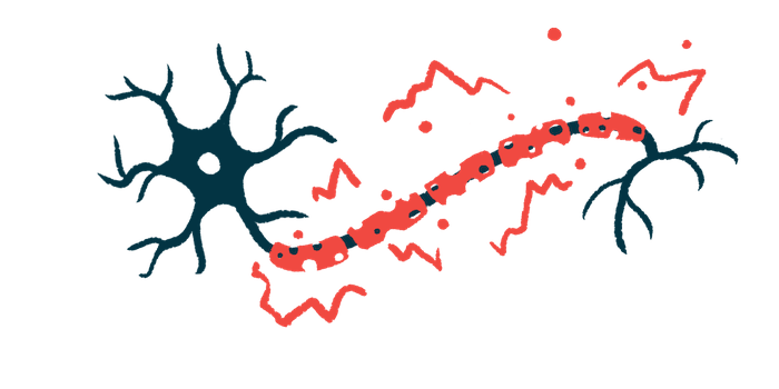
Neurofilaments are long thread-like structures that provide a framework to nerve cells, determining their shape and size. They form in the cell body and are transported down into the axon, the slender projection of nerve cells that sends out electrical signals.
NF-L is a component in neurofilaments. When nerve cells become damaged in neurodegenerative diseases such as CMT, NF-L gets shed into the bloodstream, where it can be measured as a biomarker.
A focus on glycosylation
Mutations in NEFL, the gene that provides instructions for making the NF-L protein, have been identified as the cause of CMT type 1F (CMT1F) and type 2E (CMT2E). How they affect the protein’s function is unclear, leading researchers here to focus on a type of glycosylation called O-GlcNAcylation whereby the sugar O-GlcNAc is added to proteins to shape their interactions with other proteins and regulate the way they function.
Using lab-grown human embryonic kidney cells and cells from a a cancer that forms in nerve cells, called neuroblastoma, the researchers saw that NF-L was indeed modified by O-GlcNAc, and confirmed it in samples from post-mortem brain tissue from three donors.
They then used a technique called mass spectrometry to detect glycosylated forms of NF-L and pinpoint the exact sites where O-GlcNAc was added. Five sites were identified, four in the protein’s “head” and one in its “tail.”
Individual units of NF-L are joined together in the shape of short tubes and ultimately long, or full-length, filaments. The researchers already knew the protein’s head was required for its assembly, so they mutated all of its four glycosylation sites to check how this may affect that. There was no difference in the proportion of full-length filaments between the wild-type (healthy) and mutant form.
When O-GlcNAc was induced, thus increasing global glycosylation, the proportion of full-length filaments nearly halved for the wild-type form, but not as much for the mutant form.
“These results demonstrate that site-specific O-GlcNAcylation in the NF-L head domain regulates NF assembly state,” the researchers wrote.
Effects on glycosylation
The loss of NF-L causes mitochondria, the cells’ powerhouses, to move more within nerve cells. In nerve cells from a rat hippocampus, a region in the brain, wild-type NF-L reduced the mitochondria’s movement, but the mutant form had no effect.
Glycosylation also was found to depend on the availability of nutrients, such as glucose, and to mediate interactions between individual units of NF-L as well as between NF-L and other proteins in neurofilaments.
“NF-L engages in O-GlcNAc-mediated protein-protein interactions with itself and with the NF component [alpha]-internexin, implying that O-GlcNAc may be a general regulator of NF architecture,” the researchers wrote.
Mutations in NEFL cause NF-L to build up and clump in nerve cells. To find out if they also affect glycosylation, the researchers inserted genes containing known CMT-causing mutations in human embryonic kidney cells, which then produced mutant forms of NF-L.
A total of 14 mutations were tested. Eight had abnormal glycosylation patterns compared to wild-type NF-L: six were less glycosylated than normal, and two were excessively glycosylated.
The four mutations near glycosylation sites “greatly reduced or completely abolished NF-L O-GlcNAcylation,” the researchers wrote. Moreover, these four mutant forms were insoluble, indicating they possibly clump into aggregates.
“Together, our results indicate that site-specific O-GlcNAcylation is a mode of NF regulation and may be dysregulated in neurological disorders,” the researchers wrote.
Many CMT-causing mutations resulted in abnormal glycosylation, suggesting “a potential future diagnostic benefit of characterizing NF-L O-GlcNAcylation,” they said, adding this also may be extended to other neurological disorders such as Alzheimer’s, Parkinson’s, and amyotrophic lateral sclerosis.


