AAT Protein Found to Protect Nerve Cells in CMT1A Mouse Study

Treatment with a protein called alpha-1 antitrypsin (AAT) protects nerve cells and lowers inflammation in a mouse model of Charcot-Marie-Tooth disease type 1A (CMT1A), a study has found.
Lab experiments showed that the treatment, derived from human blood (hAAT), may exert its protective effects on nerve cells by preserving myelin — the fatty protective substance surrounding nerves — and lessening inflammation.
“Given the already well-established safety profile of hAAT … we suggest that the findings of our study should be promptly investigated in CMT1A patients,” researchers wrote.
The study, “Alpha-1 Antitrypsin Reduces Disease Progression in a Mouse 2 Model of Charcot-Marie-Tooth Type 1A: A Role for Decreased Inflammation and ADAM-17 Inhibition,” was published in the International Journal of Molecular Sciences.
Like all types of Charcot-Marie-Tooth, CMT1A is a peripheral neuropathy — a disease affecting the network of nerves that mediate movements and sensations to the arms and legs.
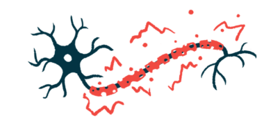
CMT1A is mainly caused by genetic changes leading to the overproduction of the PMP22 protein in Schwann cells, those responsible for producing myelin outside the brain and spinal cord. PMP22’s accumulation disrupts the structure and function of myelin, leaving nerve cells unprotected and their communication slowed.
Currently, no pharmaceutical treatments are approved specifically for CMT.
AAT is a protein elevated in the body in response to inflammation and serves to protect the body’s tissues from inflammation-induced damage. Infusions of hAAT are approved in the U.S. to treat people with AAT deficiency who have a lung condition called emphysema.
In a mouse model of multiple sclerosis, hAAT prevented myelin loss on nerve cells and lowered levels of inflammatory molecules, suggesting it could have similar effects in CMT1A.
Researchers from Switzerland and France administered the therapy in a mouse model of CMT1A to evaluate its therapeutic potential for the neuromuscular disease. Mice were given subcutaneous (under-the-skin) injections of hAAT (50 mg/kg) — or a vehicle containing no therapy — twice daily for two weeks.
What effect did the AAT protein have on CMT1A mice?
In tests of neuromuscular function, CMT1A mice fall more quickly off a rotating rod and have decreased grip strength compared with healthy mice, reflecting impaired neuromuscular function. Mice treated with hAAT showed partial, but not significant improvements in these tests, which the scientists attributed to the small number of animals used.
Consistent with neuromuscular disease, CMT1A mice also showed alterations in the electrical activity of the sciatic nerve, the body’s largest nerve that runs from the lower back down the leg. These mice have fewer nerve cell projections — called axons — in their sciatic nerve, and the axons present have a reduced diameter.
The researchers found that compound muscle action potential — the muscle response to nerve fiber stimulation — was significantly improved with hAAT treatment, with a nonsignificant increase in the number of axons present. Axon diameter was significantly improved compared to animals given the sham treatment.
Levels of the inflammatory molecule IL-6 circulating in the blood were significantly lowered with treatment, although levels of another molecule, TNF-alpha, were unchanged.
To learn more, the researchers further examined hAAT’s effects in vitro (in the lab). They found that hAAT significantly inhibited ADAM-17, a protein that acts to block myelin formation. The finding could underlie the treatment’s effects in CMT1A, the researchers noted.
In cell cultures of macrophages, a type of immune cell, that were exposed to an inflammatory substance, hAAT treatment lowered the activation of MHCII — a molecule with an important role in starting immune responses. Similar findings were observed when using a lab-made version of AAT. The treatment also led to decreases in activity levels of genes linked to inflammatory pathways.
Overall, the findings support the potential of hAAT for CMT1A, although the study was limited by its small number of animals, the researchers noted. Additional studies should expand on these findings and dive deeper into hAAT’s therapeutic mechanisms, they added.
“Our study provides initial convincing evidence for a possible role of hAAT in the management of CMT1A and possibly other peripheral neuropathies [nerve damage],” the researchers wrote, adding that it has potential to “at last offer CMT1A patients a therapeutic avenue targeting the underlying etiology [cause] of the disease rather than simply addressing symptoms that are often too divergent or broad in nature.”


